|
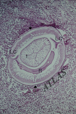
A cross section of anisakid larva shows polymyarian muscle layer, Intestine (I), Rennette cell (R), and bi-columned lateral cords (LC). Note an cellular infiltration around the larva. H & E stain.
Sung-Jong Hong
|
| CLOSE |
|
|
A cross section of anisakid larva shows polymyarian muscle layer, Intestine (I), Rennette cell (R), and bi-columned lateral cords (LC). Note an cellular infiltration around the larva. H & E stain.
Sung-Jong Hong
|
| CLOSE |